When to Stop Debriding: Clinical Signs, Risks, and Practical Guidelines for Modern Wound Care
Learn when to stop debriding in wound care. Explore clinical signs, risks, and best practices for safe, effective wound debridement management.
admin
10/29/20258 min read
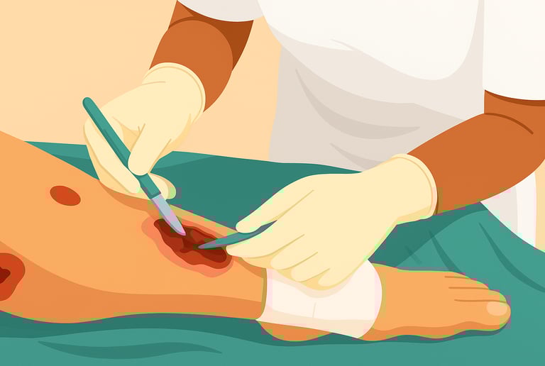
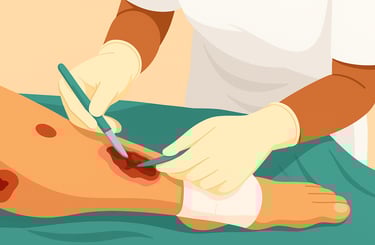
Debridement (the removal of necrotic, infected, or devitalized tissue) is a cornerstone of wound care. Performed correctly, it reduces bioburden, removes barriers to healing (slough, eschar, biofilm), and exposes healthy tissue so dressings, grafts, and other therapies can work. But debridement is not always benign; removing viable tissue, causing unnecessary bleeding or pain, or repeating aggressive procedures when they’re unlikely to help can harm the patient and delay recovery.
This guide explains when to continue debriding and, critically, when to stop. It combines guidance from professional consensus documents, systematic reviews, and clinical evidence to give clinicians practical, cautious, and actionable rules of thumb.
Quick summary
Continue debridement while the wound contains necrotic tissue, non-viable slough, heavy biofilm/bioburden, or other barriers to healing, and while the patient can tolerate further procedures.
Consider stopping or pausing debridement when: (a) you reach viable, bleeding tissue with a healthy granulation bed; (b) the patient has significant ischemia or other contraindications; (c) pain, bleeding risk, or comorbidity outweigh expected benefit; (d) repeated debridement no longer improves healing trajectory.
Use objective milestones (reduction in wound area, improved granulation, lower exudate, fewer signs of infection) and, where available, diagnostic tools (perfusion studies, infection markers, protease or POC tests) to decide about further debridement.
Below is the deeper explanation and practical algorithm.
What debridement accomplishes and why it’s usually repeated
Different debridement methods (sharp/surgical, mechanical, enzymatic, autolytic, biological/maggot therapy, hydrosurgical) share the same goal: remove devitalized tissue and disrupt barriers (necrosis, biofilm) so healing can proceed. For many chronic wounds, devitalized tissue and bioburden resurface repeatedly because the underlying cause (ischemia, venous hypertension, neuropathic pressure, malnutrition) still exists, so serial debridement is often necessary. Large observational studies and secondary analyses suggest that more frequent debridement is associated with faster healing in many wound types, which is why regular reassessment and planned repeat debridement are common practices.
However: “more frequent” is not always better for an individual patient. The clinician must balance potential benefits (cleaner bed, better perfusion of new tissue) versus harms (excess removal of viable tissue, bleeding, pain, infection spread, or destabilizing fragile tissues).
Clear reasons to stop or avoid further debridement
You have reached healthy, viable tissue that bleeds and granulates.
When sharp or surgical debridement exposes a pink, bleeding, granulating bed with advancing epithelial margins, continuing to cut risks damaging tissue that will form the new wound scaffold. In effect, stopping at a healthy margin is protective.Critical limb ischemia or inadequate perfusion is present and not corrected.
If a limb is severely ischemic (absent pedal pulses, very low ABI/toe pressures), aggressive debridement risks larger tissue loss because perfusion is insufficient to support wound repair. In ischemic wounds, coordinate with vascular specialists and consider revascularization before further aggressive debridement when possible.Patient instability or high bleeding risk.
When the patient’s hemodynamic status, anticoagulation state, platelet dysfunction, or severe comorbidities increase bleeding or procedural risk, pause or avoid surgical/sharp debridement until risks can be mitigated. Conservative or autolytic options may be safer interim choices.Pain cannot be controlled despite appropriate analgesia.
Ongoing severe pain during or after debridement that cannot be reasonably managed should prompt reconsideration of method or frequency. Techniques that reduce pain (topical anesthetic, using less aggressive modalities, or performing the procedure under regional/general anesthesia when necessary) may be preferred.No clinical benefit is seen after repeated debridement.
If serial debridements (with proper wound care and correction of underlying contributors) fail to produce improvement in key milestones (e.g., percent area reduction over 2–4 weeks, decreased exudate, better granulation) then continuing the same debridement strategy alone is unlikely to help. This is a signal to re-evaluate root causes (infection, perfusion, offloading, nutrition) or consider alternative therapies (protease modulation, NPWT, skin substitutes, or reconstructive surgery).When the tissue you would remove is viable and necessary for function.
Certain tissues (tendon, exposed bone without necrosis, viable muscle) may be only partially covered with slough but remain viable. Removing viable tissue can impair function or expose structures. In such cases, conservative, selective debridement and specialist input (orthopedics, plastic surgery) are wise.
When to pause, reassess, and change strategy
After every debridement, document the wound bed and set measurable goals. If the wound area does not improve by expected milestones (for many chronic wounds, a useful benchmark is at least 20–40% area reduction within 2–4 weeks of optimized care), pause and reassess. The literature and expert consensus emphasize using serial measurement to guide care intensity.
If bleeding is excessive or persistent, stop and manage hemostasis. Reassess coagulation status and consider alternative methods or specialist support prior to further surgery.
If infection worsens (spreading cellulitis, systemic toxicity), pause non-definitive debridement and prioritize infection control (IV antibiotics, surgical source control when indicated). Some infected wounds require staged surgical debridement in the OR rather than repeated bedside sharp debridement.
If patient-reported outcomes worsen (function, pain, quality of life) despite wound-bed changes, consider whether debridement frequency or modality is the cause and discuss alternatives with the patient and family. Shared decision-making matters.
Choosing the right debridement method — and when method affects stopping decisions
Sharp/surgical debridement is fast and often definitive. It is appropriate when large volumes of necrosis, thick eschar, or infected tissue exist and when patient condition allows. Stop when you reach bleeding viable tissue, but in some surgical scenarios staged operative debridement is planned (e.g., necrotizing infection requiring repeat OR trips).
Autolytic debridement (dressings that promote endogenous enzymatic breakdown) is selective and gentle but slower. It may be preferred when necrosis is limited or the patient cannot tolerate sharp methods. Stop when slough has separated and granulation is evident; because autolytic methods are slow, reassess frequently to avoid delaying other interventions.
Enzymatic debridement (topical proteolytic agents) is selective; stop when necrotic tissue softens and is removable. Avoid in uncontrolled infection without systemic therapy.
Biological (larval) debridement may be effective when conventional methods are unsuitable; remove maggots when necrosis is cleared or when patient intolerance occurs.
Hydrosurgical and mechanical methods are useful for specific indications; stop based on tissue appearance and hemostasis.
Regardless of method, clinicians should be prepared to switch method if progress stalls or if risks increase.
Clinical algorithm: a practical stepwise approach
Assess baseline: wound type, depth, perfusion (ABI/toe pressures), infection signs, pain, comorbidities, medications (anticoagulants), and patient preferences.
Choose an appropriate initial method (sharp for large eschar, enzymatic/autolytic for limited slough or medically fragile patients).
Debride to viable tissue — stop when a healthy, bleeding granulation bed appears or when removing further tissue would breach the structures you should preserve. Document with photos and measurements.
Set measurable milestones (area reduction percent, reduced exudate, improved granulation) and a date to reassess (commonly 1–2 weeks after intervention for chronic wounds).
If milestones met, continue planned care and consider repeating debridement at scheduled intervals if necrotic tissue recurs. If not met, re-evaluate for perfusion, infection, offloading, nutrition, protease imbalance, and consider escalating (vascular consult, infection control, protease-modulating dressings, NPWT, skin substitute, or surgery).
Special populations and caveats
Diabetic foot ulcers (DFUs): aggressive sharp debridement is often beneficial, but ischemia must be considered and vascular input sought early if perfusion is poor. Non-removable offloading and coordinated multidisciplinary care change the risk–benefit balance.
Venous leg ulcers (VLUs): debridement alone is rarely curative; compression remains the mainstay. Stop debridement when a healthy bed is present and ensure compression can be applied without harm. Evidence for debridement improving VLU healing is mixed, so balance expectations.
Cancerous or atypical wounds: certain ulcers (malignant fungating wounds, vasculitic ulcers) may need specialist input; debridement may worsen some conditions (e.g., pyoderma gangrenosum may ulcerate more with trauma), so biopsy and multidisciplinary evaluation are key before routine debridement.
Documentation, consent, and shared decision-making
Always document the indication for debridement, the method used, appearance before/after (measurements, photos), hemostasis achieved, analgesia used, and plans for reassessment. Discuss expected benefits, risks (bleeding, pain, delayed healing if viable tissue removed), and alternatives with the patient and caregivers. Shared decision-making is essential when the benefit is marginal or risks are high.
Bottom line
Debridement is a powerful and often necessary tool, but it has limits. Stop debriding when you’ve reached viable tissue, when risks (ischemia, bleeding, pain, poor perfusion) outweigh benefits, when repeated debridement does not improve healing milestones, or when specialist correction of underlying causes is needed first. Use objective milestones, multidisciplinary input, and careful documentation to guide safe decisions. Thoughtful restraint, stopping at the right time, is as important as skillful removal.
See also
Wound Debridement Guidelines: How Often Should It Be Done?
Best Practices for Chronic Wound Care: How to Assess Foot Ulcers Effectively
Why Diabetic Foot Wounds Heal Slowly: Top Factors That Delay Recovery
How Often Should Wound Dressings Be Changed? Best Practices for Healing
Best Wound Dressings for High-Exudate Wounds
More Information
For more information on the latest effective wound care, contact us to set up a time for a call.
Sources
Manna B, Nahirniak P, Morrison CA. Wound Debridement. [Updated 2023 Apr 19]. In: StatPearls [Internet]. Treasure Island (FL): StatPearls Publishing; 2025 Jan-. Available from: https://www.ncbi.nlm.nih.gov/books/NBK507882/
European Wound Management Association. Debridement: An updated overview and clarification of the principal role of debridement. Journal of Wound Care Vol 22. No 1. EWMA Document 2013. https://ewma.org/wp-content/uploads/2024/02/EWMA-Debridement-Document_JWCfinal.pdf
https://www.magonlinelibrary.com/doi/abs/10.12968/jowc.2013.22.Sup1.S1Wilcox JR, Carter MJ, Covington S. Frequency of Debridements and Time to Heal: A Retrospective Cohort Study of 312 744 Wounds. JAMA Dermatol. 2013;149(9):1050–1058. doi:10.1001/jamadermatol.2013.4960 https://jamanetwork.com/journals/jamadermatology/fullarticle/1720508
https://pubmed.ncbi.nlm.nih.gov/23884238/Best practice for wound debridement (International Consensus Document). Journal of Wound Care. Vol 33. No 6. Sup C. June 2024. https://www.journalofwoundcare.com/docs/debridement-consensus.pdf. journalofwoundcare.com
Nowak M, Mehrholz D, Barańska-Rybak W, Nowicki RJ. Wound debridement products and techniques: clinical examples and literature review. Postepy Dermatol Alergol. 2022 Jun;39(3):479-490. doi: 10.5114/ada.2022.117572. Epub 2022 Jul 14. PMID: 35950126; PMCID: PMC9326937. https://pmc.ncbi.nlm.nih.gov/articles/PMC9326937/
Elraiyah T., et al. A systematic review and meta-analysis of debridement methods for chronic diabetic foot ulcers. Journal of Vascular Surgery. 2016. doi: 10.1016/j.jvs.2015.10.002 https://www.jvascsurg.org/article/S0741-5214(15)02024-8/fulltext
Gethin G, Cowman S, Kolbach DN. Debridement for venous leg ulcers. Cochrane Database of Systematic Reviews 2015, Issue 9. Art. No.: CD008599. DOI: 10.1002/14651858.CD008599.pub2. https://www.cochrane.org/evidence/CD008599_debridement-venous-leg-ulcers
Deena Clare Thomas, Chong Li Tsu, Rose A. Nain, Norkiah Arsat, Soong Shui Fun, Nik Amin Sahid Nik Lah,
The role of debridement in wound bed preparation in chronic wound: A narrative review, Annals of Medicine and Surgery, Volume 71, 2021, 102876, ISSN 2049-0801, https://doi.org/10.1016/j.amsu.2021.102876
https://www.sciencedirect.com/science/article/pii/S2049080121008268
Journal of Community Nursing. Wound Debridement Guidelines and Practice to Remove Barriers to Healing. https://www.jcn.co.uk/uploads/resources/62d1283436aeae1ec417f889360fc32a.pdf
"Wound Debridement." Physiopedia, . 24 Oct 2020, 13:24 UTC. 29 Oct 2025, 18:07 https://www.physio-pedia.com/index.php?title=Wound_Debridement&oldid=257226
Southwest Regional Wound Care Program. Guideline and Procedures: Wound Debridement (excluding conservative sharp debridement). 2015. https://www.swrwoundcareprogram.ca/uploads/contentdocuments/wounddebridement.pdf. swrwoundcareprogram.ca
Wormald JCR, Wade RG, Dunne JA, Collins DP, Jain A. Hydrosurgical debridement versus conventional surgical debridement for acute partial‐thickness burns. Cochrane Database of Systematic Reviews 2020, Issue 9. Art. No.: CD012826. DOI: 10.1002/14651858.CD012826.pub2. https://www.cochranelibrary.com/cdsr/doi/10.1002/14651858.CD012826.pub2/full
Hinchliffe RJ, Forsythe RO, Apelqvist J, et al. Guidelines on diagnosis, prognosis, and management of peripheral artery disease in patients with foot ulcers and diabetes (IWGDF 2019 update). Diabetes Metab Res Rev. 2020;36(S1):e3276. https://doi.org/10.1002/dmrr.3276 https://iwgdfguidelines.org/wp-content/uploads/2020/11/Hinchliffe_et_al-2020-IWGDF-PAD-guideline.pdf
International Wound Infection Institute. WOUND INFECTION IN CLINICAL PRACTICE, Principles of best practice. 2022. https://woundinfection-institute.com/wp-content/uploads/IWII-CD-2022-web-1.pdf
Westby MJ, Dumville JC, Stubbs N, Norman G, Wong JK, Cullum N, Riley RD. Protease activity as a prognostic factor for wound healing in venous leg ulcers. Cochrane Database Syst Rev. 2018 Sep 1;9(9):CD012841. doi: 10.1002/14651858.CD012841.pub2. PMID: 30171767; PMCID: PMC6513613. https://www.ncbi.nlm.nih.gov/pmc/articles/PMC6513613/
* This blog is for informational purposes only and is not a substitute for professional medical advice, diagnosis, or treatment.