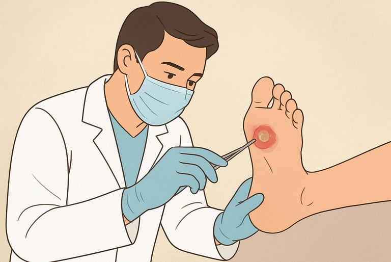
Best Practices for Chronic Wound Care: How to Assess Foot Ulcers Effectively
Learn best practices for chronic wound care. Discover how to assess diabetic foot ulcers effectively with proven tools, tips, and clinical guidance
admin
9/19/20256 min read


Why systematic assessment matters in chronic wound care
A comprehensive foot ulcer assessment reduces the risk of complications like infection, gangrene, and amputation. It helps clinicians prioritize urgent problems (ischemia, deep infection, spreading cellulitis) and choose targeted interventions such as revascularization, debridement, offloading, or advanced therapies (skin substitutes, negative pressure wound therapy). The International Working Group on the Diabetic Foot (IWGDF) and other clinical reviews advocate using structured assessment pathways and early multidisciplinary referral when indicated.
Quick checklist: the core components of a foot ulcer assessment
Patient history (medical, social, medication).
Wound history (onset, duration, prior treatments, response).
Wound inspection (location, size, depth, wound bed, exudate, odor).
Vascular assessment (pulses, ABI, toe pressures, TcPO₂ when needed).
Neurologic assessment (protective sensation testing).
Infection assessment (local and systemic signs; probe-to-bone test).
Classification/scoring (PEDIS, SINBAD, Wagner, others).
Photodocumentation and serial measurement.
Treatment plan (offloading, debridement, dressings, antibiotics, referral).
Using this checklist consistently improves documentation, communication, and healing tracking. Several validated classification and scoring systems (PEDIS, SINBAD, Wagner) help stratify risk and guide treatment decisions.
1. Thorough history: start with risk and context
Collect a focused history every time. Key items:
Diabetes control (A1c if available), duration of diabetes, recent glucose trends.
Peripheral arterial disease (claudication, prior vascular procedures).
Neuropathy symptoms (burning, numbness, tingling).
Prior foot ulcers, prior amputations or revascularization.
Current medications (especially immunosuppressants, steroids, SGLT2 inhibitors discussions where relevant).
Social context (mobility, footwear, ability to offload, home support).
History shapes urgency: a new ulcer in a patient with rest pain or prior bypass merits rapid vascular workup; neuropathic insensate feet require strict offloading and patient education. Evidence-based guidelines emphasize early risk stratification and regular screening for high-risk patients.
2. Wound inspection: what to look for and how to record it
Use a systematic inspection and document:
Location: plantar, dorsal, toes, heel - location affects offloading strategy.
Size and shape: measure length × width (cm) and photograph with a scale. Consistent measurement technique improves tracking of wound area over time.
Depth and tissue types: note presence of slough, granulation tissue, exposed tendon or bone.
Exudate and odor: amount, character (serous, purulent), and odor can suggest infection or necrosis.
Periwound skin: maceration, callus, erythema, induration. Callus often indicates pressure - offloading needed.
Foreign bodies and undermining/tunneling: probe gently to map tunnels.
Photodocumentation and digital wound measurement tools are helpful for serial monitoring, but ensure consistent lighting, distance, and scale. Wound bed preparation principles apply: assess for devitalized tissue that may require debridement, and for healthy granulation that indicates progress.
3. Vascular assessment: don’t miss ischemia (peripheral arterial disease)
Ischemia significantly impairs healing. Components of vascular testing include:
Palpation of pedal pulses (dorsalis pedis, posterior tibial). Absent or diminished pulses should prompt further testing.
Ankle-brachial index (ABI): a first-line noninvasive test but can be falsely elevated in calcified arteries (common in diabetes).
Toe pressures and toe–brachial index (TBI): useful when ABI is unreliable; toe pressures <30–50 mmHg often indicate poor healing potential.
Transcutaneous oxygen pressure (TcPO₂): sometimes used to predict healing and revascularization benefit.
Duplex ultrasound / arterial imaging: used when revascularization is being considered.
Early vascular assessment and timely vascular referral for revascularization (when indicated) are recommended by diabetic foot guidelines because restoring perfusion often determines healing potential. Avoid making assumptions about perfusion based on appearance alone. Measure.
4. Neurologic testing: detect loss of protective sensation
Loss of protective sensation (LOPS) is a key risk for ulceration and recurrence. Use simple, validated tests:
10-gram monofilament testing at standardized points on the plantar surface. Inability to feel the monofilament suggests LOPS.
Vibration testing with a 128-Hz tuning fork can complement monofilament testing.
Document sensory testing results and integrate them into patient education and offloading decisions. Patients with neuropathy need regular exam intervals and footwear interventions to reduce pressure-related ulcers.
5. Infection assessment: recognize local and systemic signs
Infection can be limb- or life-threatening. Key steps:
Look for local signs: increasing erythema, warmth, purulent discharge, induration, foul odor, newly increased pain (if sensation present).
Look for systemic signs: fever, tachycardia, hypotension, leukocytosis - these warrant urgent care.
Use the probe-to-bone test (gently probing the ulcer to see if bone can be contacted) as an inexpensive clinical tool that, if positive, increases suspicion for osteomyelitis and the need for imaging and further workup.
Obtain wound cultures appropriately - deep tissue or bone cultures are preferred when available; superficial swabs are less reliable for guiding therapy.
Guidelines recommend classifying infection severity and treating promptly; severe or rapidly progressing infections require hospitalization and multidisciplinary management. Avoid overuse of antibiotics for noninfected wounds; base antibiotic decisions on infection signs and cultures.
6) Use structured classification systems for consistency
Several validated classification tools help standardize assessment and predict outcomes:
PEDIS (Perfusion, Extent/size, Depth/tissue loss, Infection, Sensation) — useful for comprehensive grading. Studies report good reliability and clinical utility.
SINBAD (Site, Ischemia, Neuropathy, Bacterial infection, Area, Depth) — a simple scoring system validated for outcome prediction in some cohorts.
Wagner classification — widely used; simple but focuses mainly on depth and gangrene and is less granular for infection/perfusion.
No single system is perfect; choose one that fits your practice and use it consistently to monitor healing trajectories and support referrals. Comparative reviews recommend using structured scores to improve communication and patient triage.
7) Imaging and advanced diagnostics: when and what to order
Plain radiographs (X-ray): first-line for suspected osteomyelitis, gas in tissues, foreign bodies, or bony deformities; radiographic changes lag behind clinical infection.
MRI: best for soft tissue and bone infection (osteomyelitis) when diagnosis is uncertain.
Bone scan / nuclear imaging: alternative when MRI contraindicated.
Vascular imaging: duplex ultrasound, CT angiography, or MR angiography for ischemia evaluation.
Order imaging driven by clinical suspicion, especially when probe-to-bone is positive, infection is suspected, or ischemia limits healing.
8. Documentation, serial measurement, and photography
High-quality documentation supports clinical decisions, billing, and outcome tracking:
Use consistent measurement methods and wound area calculations (length × width or digital planimetry).
Time-stamped photographs with a measurement scale standardize comparisons.
Document key elements: size, depth, tissue types, exudate, vascular and sensory findings, and applied interventions (debridement, offloading device, antibiotics).
Regular reassessment (weekly or as clinically appropriate) helps detect deterioration early and shows progress or lack thereof to guide escalation. Evidence suggests that early improvement (e.g., ≥50% area reduction by 4 weeks) correlates with eventual healing, which can inform decisions about advanced therapies.
9. Translating assessment into action: typical next steps
Ischemia suspected → vascular testing and rapid referral for revascularization evaluation.
Infection suspected → appropriate cultures, imaging, start empiric antibiotics for moderate–severe infections and consider hospitalization for systemic signs.
Neuropathic, noninfected ulcer → offloading (total contact cast, removable walker, or appropriate footwear), sharp debridement, moist wound dressings, and patient education.
Nonhealing despite basic care → consider advanced therapies (negative pressure wound therapy, skin substitutes, growth factors) and multidisciplinary review. Evidence for advanced modalities varies; consider specialist input and review current guidelines and local formularies.
Red flags and reasons for urgent referral
Refer urgently (same day or within 24 hours) for: spreading cellulitis, suspected necrotizing infection, systemic sepsis signs, critical limb ischemia (rest pain, tissue loss with severe ischemia), exposed bone with suspected osteomyelitis, rapidly progressing gangrene. Early multidisciplinary involvement (vascular surgery, infectious disease, podiatry, wound care specialists) improves outcomes in complex cases.
Communication, patient education, and prevention
Assessment is only valuable if it leads to education and behavior change:
Teach daily foot inspection, proper footwear and sock selection, and daily moisturizing for dry skin (avoid between toes).
Stress the importance of offloading and activity modification while a wound heals.
Schedule regular follow-up and screening for high-risk patients to prevent recurrence. Preventive strategies and patient empowerment are cornerstones of long-term chronic wound care.
Limitations
Clinical judgment should guide every assessment. Tools and scores assist but do not replace individualized care. Some tests (ABI) can be misleading in people with arterial calcification; imaging and toe pressures may be needed. The evidence base for certain advanced therapies is evolving. Refer to current guidelines and recent systematic reviews before widespread adoption. The recommendations here are intended to be practical and cautious rather than absolute.
See Also
How to Tell If a Wound Is Healing: Signs of Proper Wound Care Progress
More information
For more information on the latest effective wound care, contact us to set up a time for a call.
Sources
NICE guideline NG19 — Diabetic foot problems: prevention and management. (UK guideline; updated surveillance)
Armstrong DG, et al. Diabetic foot ulcers: a review. (2023) — clinical overview on assessment and management principles. PubMed
Chuan F., et al. Reliability and validity of the PEDIS classification system. PLoS ONE (2015) — evidence for PEDIS utility. PLOS One
Monteiro-Soares M., et al. Guidelines on the classification of diabetic foot ulcers. (2020) — review of classification systems (PEDIS, SINBAD, Wagner). PubMed
IWGDF / IDSA updates on diabetic foot infection diagnosis and management (2023/2024 updates).
New Evidence-Based Therapies for Complex Diabetic Foot Wounds - American Diabetes Association
* This blog is for informational purposes only and is not a substitute for professional medical advice, diagnosis, or treatment.
