When to Use Skin Substitutes or Grafts for Non-Healing Wounds
Learn when to use skin substitutes or grafts for non-healing wounds with evidence-based wound care practices to enhance healing and patient outcomes.
admin
10/18/20258 min read
Non-healing wounds—whether diabetic foot ulcers, venous leg ulcers, or complex surgical defects—are a major clinical challenge. When standard wound care (debridement, infection control, offloading, compression, moisture balance) does not produce steady progress, clinicians may consider skin substitutes, grafts, and cellular/tissue-based products as part of a strategy to promote closure and restore durable skin coverage. This article explains when these options are appropriate, what types exist, how to choose between them, and practical considerations for clinicians.
Quick summary
Consider skin substitutes or grafts when a wound has failed to progress after optimal standard care, and key barriers (infection, ischemia, offloading, nutrition) have been addressed.
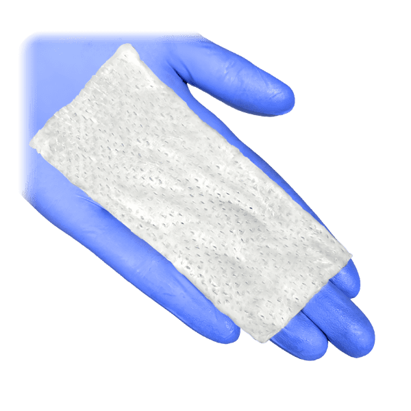
Different products suit different wounds: split-thickness skin grafts may be best for well-vascularized beds; acellular dermal matrices and bi-layer templates (e.g., Integra, Coll-e-Derm™, Resolve Matrix™) help with complex wounds and exposed structures; cellular or placental-derived products (e.g., Dermagraft, Apligraf, EpiFix, Restorigin™) are used for chronic ulcers that need biologic stimulation.
Evidence shows many skin substitutes increase healing rates compared with standard care in the short term, but long-term outcomes, recurrence, and cost-effectiveness vary. Use shared decision-making.
1. First ask: has standard care been optimized?
Before applying skin substitutes or grafts, verify that common, modifiable barriers to healing have been addressed. These include:
Infection control: Active infection should be treated per guidelines; a clean wound bed is more likely to accept grafts or substitutes.
Perfusion: Assess for peripheral arterial disease; revascularization may be necessary before grafting in ischemic limbs.
Mechanical factors: Offloading (for plantar ulcers) and pressure redistribution must be optimized.
Debridement and wound bed preparation: Necrotic tissue and biofilm can hinder engraftment—adequate debridement and moisture balance matter.
Metabolic and systemic factors: Glycemic control, nutrition, and smoking cessation should be considered as part of the plan.
If these elements are not in place, advanced products are less likely to succeed. The IWGDF wound-healing guidelines recommend addressing these core issues before escalation.
2. When to consider skin substitutes or grafts
You may consider tissue replacement when one or more of the following apply:
A chronic wound has failed to achieve meaningful progress with standard care over an evidence-based time frame (often several weeks to months depending on wound type and severity).
Wound depth or tissue loss requires structural reconstruction (exposed tendon, bone, or deep soft tissue defects) where dermal templates or grafts can provide coverage and scaffold.
Recurrent or stalled ulcers despite offloading and optimization where biologic stimulation (cellular products or placental grafts) may help achieve closure.
Patient factors and goals (for example, a patient who prioritizes limb salvage and is willing/able to adhere to follow-up and offloading may be a candidate for advanced products).
Clinical judgment and multidisciplinary input (podiatry, vascular surgery, wound care specialists) are recommended when escalation is considered.
3. Types of skin substitutes and grafts
There are many products marketed as skin substitutes or tissue-based products. Clinicians should understand basic categories:
A. Autografts (split-thickness skin grafts, aka STSG)
Use the patient’s own skin; require a well-vascularized wound bed to survive. Often used for larger skin defects after adequate debridement. They provide durable coverage but require a donor site and are operator-dependent. Evidence supports use in appropriately prepared, perfused beds.
B. Acellular dermal matrices and bilayer templates (e.g., Integra, Coll-e-Derm™, Resolve Matrix™)
Provide a collagen/dermal scaffold that supports revascularization and cellular infiltration; often used when soft tissue is lost or when covering exposed structures. May be paired with later STSG. Outcomes are generally favorable for complex wounds, though technique and patient selection matter.
C. Cellular and tissue-based products (CTPs) — living or cellular products (e.g., Dermagraft, Apligraf)
Contain viable cells or living dermal constructs intended to deliver growth factors and cytokines. RCTs have shown improved closure rates for some products in diabetic foot ulcers. These are typically applied to chronic wounds that have a clean bed but are stalled in healing.
D. Placental/amnion-derived products (e.g., dehydrated human amniotic membrane, EpiFix, Restorigin™)
Placental tissues provide extracellular matrix components, growth factors, and anti-inflammatory properties. Systematic reviews suggest benefit for some chronic wounds, including DFUs, though study designs vary.
E. Xenografts and bioscaffolds (e.g., SIS, porcine small intestinal submucosa)
These are biologic scaffolds used for structural support and tissue ingrowth in select defects. Evidence varies by product and indication.
4. Matching product to wound
Choosing the right option depends on wound characteristics, host factors, and resources:
Is the wound bed well vascularized and free of infection? If yes, STSG or cellular products may be reasonable. If no, address ischemia/infection first.
Is there exposed tendon, bone, or hardware? Bilayer dermal templates (Integra) or staged reconstruction may be preferred before a skin graft.
Is the wound chronic but superficial and clean? Placental or cellular products (Restorigin™, EpiFix, Apligraf, Dermagraft) have RCT and real-world evidence suggesting improved closure rates when used alongside standard care.
Patient adherence and follow-up: Some grafts and substitutes require strict offloading and wound protection after application; the patient’s ability to comply is crucial.
Cost and payer policy also affect choice. Some products are expensive and may require prior authorization; check local coverage policies and evidence for the selected product.
5. Evidence summary: what the literature suggests
Many skin substitutes increase short-term healing rates compared with standard care in DFUs and some chronic wounds. Systematic reviews and meta-analyses support an effect on closure at defined endpoints. Yet long-term data on recurrence, limb salvage, and cost-effectiveness are more limited.
Specific RCTs (Dermagraft, Apligraf, EpiFix) have shown improved closure for selected patients when used with standard care. Results vary by patient selection, wound age, and protocol.
Placental/amnion products show promise in multiple observational studies and some controlled trials; systematic reviews suggest benefit but call for higher-quality, long-term studies.
In short: evidence supports selective use of these products in appropriate patients, but outcomes depend on context and adjunctive care.
6. Practical protocol for clinics
Optimize standard care (infection control, perfusion, offloading, debridement, nutrition).
Risk-stratify the wound (depth, exposed structures, duration, prior recurrence).
Discuss options in a multidisciplinary setting when possible.
Select product based on wound biology and patient factors (see section 4).
Ensure patient understands post-procedure care (offloading, wound protection, follow-up visits).
Measure outcomes (time to closure, durability, recurrence, adverse events) and document for quality improvement.
7. Risks, limitations, and cost considerations
Not a panacea: Advanced products help many wounds heal faster, but they do not replace the need to fix underlying problems (ischemia, pressure, infection).
Variable evidence for long-term outcomes: Fewer high-quality trials report long-term recurrence or limb salvage benefits. Cost-effectiveness is product- and setting-dependent.
Access and payer rules: Reimbursement varies; clinicians should check local coverage and prepare documentation that justifies product use.
8. Case examples
Case A — chronic DFU, clean base, neuropathic, good perfusion: Consider a cellular product (e.g., Dermagraft) or placental allograft (e.g., Restorigin™) adjunct to continued offloading and standard care. Monitor closure over recommended timeframes.
Case B — large post-debridement defect with exposed tendon but good perfusion: A dermal regeneration template (e.g., Coll-e-Derm™) followed by STSG may provide best structural outcome.
Case C — recurrent venous leg ulcer: Evaluate compression therapy and venous disease; if standard care optimized but stalled, consider skin grafting or tissue replacement in select patients with appropriate vascular status. Evidence supports grafting in some venous ulcer cases, but selection and technique matter.
9. Documentation and shared decision-making
Document the wound-care pathway, rationale for using an advanced product, expected benefits, alternatives, and the post-application plan (offloading, dressing schedule, follow-up cadence). Discuss costs and uncertainties with the patient and family. This transparency supports ethical, patient-centered care.
Conclusion
Skin substitutes, grafts, and cellular/tissue-based products are useful tools for selected non-healing wounds. They are most effective when used after standard care optimization and in the right wound context: clean bed, adequate perfusion, controlled infection, and reliable offloading or compression when indicated. Evidence supports improved short-term healing with several products, but long-term benefits, recurrence reduction, and cost-effectiveness require further high-quality study. Use multidisciplinary evaluation, tailor the choice of product to wound biology and patient circumstances, and apply shared decision-making to align treatment with patient goals.
See also
Best Practices for Chronic Wound Care: How to Assess Foot Ulcers Effectively
How to Tell If a Wound Is Healing: Signs of Proper Wound Care Progress
Why Diabetic Foot Wounds Heal Slowly: Top Factors That Delay Recovery
How Often Should Wound Dressings Be Changed? Best Practices for Healing
Wound Care Guide: How to Tell Colonization from True Infection
More Information
For more information on the latest effective wound care, contact us to set up a time for a call.
Sources
Chen P, et al. Guidelines on interventions to enhance healing of foot ulcers in people with diabetes (2023). International Working Group on the Diabetic Foot (IWGDF). https://iwgdfguidelines.org/wp-content/uploads/2023/07/IWGDF-2023-07-Wound-Healing-Guideline.pdf
Santema TB, Poyck PPC, Ubbink DT. Skin grafting and tissue replacement for treating foot ulcers in people with diabetes. Cochrane Database of Systematic Reviews 2016, Issue 2. Art. No.: CD011255. DOI: 10.1002/14651858.CD011255.pub2. https://www.cochranelibrary.com/cdsr/doi/10.1002/14651858.CD011255.pub2/full
Santema TB, Poyck PP, Ubbink DT. Systematic review and meta-analysis of skin substitutes in the treatment of diabetic foot ulcers: Highlights of a Cochrane systematic review. Wound Repair and Regeneration. 2016 Jul;24(4):737-44. doi: 10.1111/wrr.12434. Epub 2016 Jun 3. PMID: 27062201.
https://pubmed.ncbi.nlm.nih.gov/27062201/William A. Marston, Jason Hanft, Paul Norwood, Richard Pollak, for the Dermagraft Diabetic Foot Ulcer Study Group; The Efficacy and Safety of Dermagraft in Improving the Healing of Chronic Diabetic Foot Ulcers: Results of a prospective randomized trial. Diabetes Care 1 June 2003; 26 (6): 1701–1705. https://doi.org/10.2337/diacare.26.6.1701. https://diabetesjournals.org/care/article/26/6/1701/26323/The-Efficacy-and-Safety-of-Dermagraft-in-Improving
Edmonds M. Apligraf in the Treatment of Neuropathic Diabetic Foot Ulcers. The International Journal of Lower Extremity Wounds. 2009;8(1):11-18. doi:10.1177/1534734609331597 https://pubmed.ncbi.nlm.nih.gov/19189997/. https://journals.sagepub.com/doi/10.1177/1534734609331597
Lakmal K, Basnayake O, Hettiarachchi D. Systematic review on the rational use of amniotic membrane allografts in diabetic foot ulcer treatment. BMC Surgery. 2021 Feb 15;21(1):87. doi: 10.1186/s12893-021-01084-8. PMID: 33588807; PMCID: PMC7885244. https://pubmed.ncbi.nlm.nih.gov/33588807/
Mohammed YA, Farouk HK, Gbreel MI, Ali AM, Salah AA, Nourelden AZ, Gawad MMA. Human amniotic membrane products for patients with diabetic foot ulcers. do they help? a systematic review and meta-analysis. Journal of Foot and Ankle Research. 2022 Sep 14;15(1):71. doi: 10.1186/s13047-022-00575-y. PMID: 36104736; PMCID: PMC9472416. https://pmc.ncbi.nlm.nih.gov/articles/PMC9472416/
Chang DK, Louis MR, Gimenez A, Reece EM. The Basics of Integra Dermal Regeneration Template and its Expanding Clinical Applications. Seminars in Plastic Surgery. 2019 Aug;33(3):185-189. doi: 10.1055/s-0039-1693401. Epub 2019 Aug 2. PMID: 31384234; PMCID: PMC6680073. https://www.ncbi.nlm.nih.gov/pmc/articles/PMC6680073/
Giovannini UM, Teot L. Long-term follow-up comparison of two different bi-layer dermal substitutes in tissue regeneration: Clinical outcomes and histological findings. International Wound Journal. 2020 Oct;17(5):1545-1547. doi: 10.1111/iwj.13381. Epub 2020 May 2. PMID: 32359006; PMCID: PMC7948946. https://www.ncbi.nlm.nih.gov/pmc/articles/PMC7948946/
Zelen CM, Gould L, Serena TE, Carter MJ, Keller J, Li WW. A prospective, randomised, controlled, multi-centre comparative effectiveness study of healing using dehydrated human amnion/chorion membrane allograft, bioengineered skin substitute or standard of care for treatment of chronic lower extremity diabetic ulcers. International Wound Journal. 2015 Dec;12(6):724-32. doi: 10.1111/iwj.12395. Epub 2014 Nov 26. PMID: 25424146; PMCID: PMC7950807. https://pubmed.ncbi.nlm.nih.gov/25424146/
Centers for Medicare & Medicaid Services — Skin Substitute Grafts/Cellular and Tissue-Based Products for the Treatment of Diabetic Foot Ulcers and Venous Leg Ulcers.
https://www.cms.gov/medicare-coverage-database/view/lcd.aspx?lcdid=39870&ver=4Jones JE, Nelson EA, Al‐Hity A. Skin grafting for venous leg ulcers. Cochrane Database of Systematic Reviews 2013, Issue 1. Art. No.: CD001737. DOI: 10.1002/14651858.CD001737.pub4. https://www.cochranelibrary.com/cdsr/doi/10.1002/14651858.CD001737.pub4/full
Santema TB, Poyck PP, Ubbink DT. Systematic review and meta-analysis of skin substitutes in the treatment of diabetic foot ulcers: Highlights of a Cochrane systematic review. Wound Repair and Regeneration. 2016 Jul;24(4):737-44. doi: 10.1111/wrr.12434. Epub 2016 Jun 3. PMID: 27062201. https://pubmed.ncbi.nlm.nih.gov/27062201/
* This blog is for informational purposes only and is not a substitute for professional medical advice, diagnosis, or treatment.