Wound Debridement Guidelines: How Often Should It Be Done?
Learn how often to perform wound debridement for optimal healing. Explore best practices, methods, and guidelines for effective wound care management.
admin
10/14/20258 min read
Debridement, the removal of devitalized (dead) tissue, foreign material, and excessive biofilm from a wound, is a fundamental step in wound bed preparation. Done well, it improves the wound environment, reduces bacterial load, helps dressings contact healthy tissue, and supports the normal phases of healing. But clinicians and caregivers frequently ask: how often should debridement be performed? The short answer is “it depends” on wound type, amount of necrosis or slough, infection status, healing trajectory, patient comorbidities, and the debridement method used. This article lays out practical, evidence-informed guidance on frequency, methods, monitoring, and when to change course.
Why debridement frequency matters
Debridement is an active intervention. Each episode changes the wound surface and the microbiologic environment. Too-frequent aggressive debridement may damage viable tissue, increase bleeding risk, or increase pain; too-infrequent debridement allows necrotic tissue, biofilm, and proteases to persist, which can stall healing and increase infection risk. The goal is to remove non-viable tissue often enough to keep the wound progressing, but not so often that you cause harm. Major wound-care guidance documents and consensus statements stress regular, individualized reassessment rather than a fixed universal schedule.
Common debridement methods (and how frequency differs)
Different debridement techniques have different optimal frequencies and practical constraints. Knowing how each method works helps you decide how often to use it.
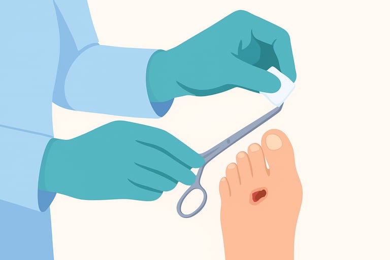
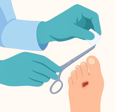
Conservative sharp wound debridement (CSWD) - selective removal of slough and callus using scalpel/scissors at the bedside. Often performed at every clinic visit if there is recurrent slough or callus; many podiatrists and wound clinics report weekly or every-other-week schedules in routine practice. CSWD is efficient but requires trained clinicians and judgement about depth.
Surgical (operating room) debridement - aggressive excision used for extensive necrosis, deep infection, or when large volumes of tissue are unstable. Frequency depends on clinical course; often a single definitive debridement followed by reassessment, with repeat procedures if infection or necrosis recurs.
Autolytic debridement - uses the body’s own enzymes and a moist dressing (e.g., hydrogel, hydrocolloid) to soften necrotic tissue. This is the gentlest method but can be slower and often requires daily to several-day dressing management; clinician review every 3–7 days is common.
Enzymatic debridement - topical proteolytic agents (e.g., collagenase) applied per product instructions; dressings typically changed per manufacturer guidance (often every 24–72 hours) and clinician reassessment occurs regularly (weekly or sooner if infection).
Maggot (larval) debridement therapy (MDT) - medical-grade larvae selectively remove devitalized tissue; typical treatment cycles last several days with repeats as needed and clinician monitoring at intervals recommended in protocols. MDT often works quickly for sloughy wounds and is considered when other methods are inappropriate.
Low-frequency ultrasound debridement (LFUD) - device-based cavitation that helps remove slough and biofilm; some studies show LFUD used 2–3 times per week may be effective, although protocols vary.
Hydrosurgical and pulsed lavage - higher-pressure mechanical cleansing used in the OR or procedure room; frequency depends on how much devitalized tissue is present and the clinical response.
Evidence on frequency: what studies and surveys show
There is not a universal, high-quality randomized trial that sets one frequency for all wounds and methods. Instead, the evidence base offers patterns and practical guidance:
Surveys and audits of routine practice show many clinicians perform conservative sharp debridement at weekly or every two weeks intervals for chronic foot ulcers and other wounds, with variability by clinician, country, and setting. A cross-sectional study of podiatrists reported CSWD performed at every visit by most clinicians, with typical intervals of weekly or every two weeks.
A randomized trial comparing weekly debridement vs. every-second-week debridement found that standardized care including either weekly or fortnightly debridement could achieve good healing rates. But both schedules were used within a broader package of care; the study suggested that a range of frequencies may be reasonable when applied in the right clinical context. This highlights that frequency interacts with overall wound management (dressings, offloading, infection control).
Systematic reviews emphasize that frequent debridement is often associated with improved wound bed preparation and may accelerate healing in some wound types, but study heterogeneity limits definitive, one-size-fits-all recommendations. Many guideline documents therefore recommend individualized, frequent reassessment.
For device-based debridement such as low-frequency ultrasound, smaller trials suggest thrice-weekly treatments may be effective for sloughy, chronic wounds, but evidence is still emerging and protocols vary.
Practical, evidence-informed frequency recommendations
Below are pragmatic starting points that align with guidelines, expert consensus, and common practice. Use them as frameworks, not rigid rules.
Initial assessment and early phase (first 1–2 weeks)
If substantial necrosis, slough, callus, or biofilm is present, consider surgical or sharp debridement promptly to convert to a clean wound bed. Early, decisive debridement can remove infection niduses and speed recovery. After initial aggressive debridement, plan close follow-up (often weekly) to reassess.
Ongoing conservative sharp debridement (CSWD)
When CSWD is the chosen method (for example, in many diabetic foot clinics), expect weekly to fortnightly sessions depending on reaccumulation of slough, callus formation, exudate level, and wound progress. If slough or callus re-forms rapidly, weekly debridement may be needed. If the wound is clearly improving and tissue remains healthy, lengthen intervals.
Autolytic or enzymatic debridement
Use these gentler methods when selective debridement is needed, for painful wounds, or when vascular supply is limited. Dressings and topical enzymes typically require daily to every-few-day dressing changes; clinician reassessment every 1–2 weeks is common to ensure progress.
Device-assisted methods (LFUD, NPWT with irrigation, hydrosurgery)
Follow device protocols; LFUD may be used 2–3 times per week, NPWT reduces need for frequent dressing changes and thus surgical debridement frequency, and hydrosurgery is used as indicated (often as single-session OR procedures with reassessment).
Infected wounds or osteomyelitis
If infection is present, combine debridement with systemic antibiotics and more frequent reassessment (often multiple times per week initially). Surgical debridement may need repeating until infection is controlled.
Maintenance phase
Once a wound has a healthy, stable granulation bed and epithelialization is progressing, debridement frequency may be reduced. Continue to remove minor slough or callus that could interfere with closure This may be every 1–4 weeks depending on reaccumulation.
Clinical signs that indicate more frequent debridement is needed
Rapid re-accumulation of slough or callus.
Persistent or increasing wound exudate, malodor, or visible biofilm.
Failure to form healthy granulation tissue despite other optimized care.
Clinical or laboratory markers suggesting ongoing infection.
High-risk anatomic sites (e.g., weight-bearing plantar ulcers with recurrent callus).
If you observe any of these, escalate debridement frequency and reassess the overall plan; address perfusion, offloading, glycemic control, nutrition, and infection.
Safety, documentation, and team roles
Who should debride? Methods vary in required expertise. Surgical debridement belongs to surgeons; CSWD is typically done by trained podiatrists, wound specialists, or nurses following local scope-of-practice rules; autolytic and enzymatic methods can be managed by trained clinicians and sometimes caregivers under guidance. Major guidance documents emphasize trained personnel and multidisciplinary involvement.
Documentation: At each debridement, document wound size, tissue types removed, bleeding, pain, and whether deeper structures were exposed. This supports decisions about frequency, reimbursement, and safety. CMS and local payers also require reasonable medical necessity and documentation when billing multiple debridements.
Pain and hemostasis: Manage pain (topical or local anesthetic as appropriate) and be prepared to stop debridement if bleeding or patient distress occurs. Repeat debridement is not routine if hemostasis cannot be achieved safely.
Putting frequency into a broader pathway (TIMERS and wound bed preparation)
Debridement is one step in an overall wound-care pathway (e.g., TIMERS: Tissue management, Inflammation/infection control, Moisture balance, Edge of wound/epithelialization, Repair/regeneration, Social factors). Frequency decisions should be integrated into this whole-patient approach: if debridement alone does not produce progress, reassess systemic factors (perfusion, nutrition, glycemic control), offloading, and the need for advanced therapies.
Practical clinic checklist for deciding debridement frequency
Is there devitalized tissue, thick slough, or callus impairing the wound? → debride.
Was the wound infected or at high infection risk? → combine debridement with antibiotics and reassess more often.
After debridement, is healthy granulation forming within 1–2 weeks? → maintain or lengthen intervals.
Is slough/callus returning rapidly? → increase frequency (weekly) or consider different method.
Is the patient at bleeding risk or intolerant to sharp debridement? → prefer gentler autolytic/enzymatic or MDT, with close follow-up.
Limitations
High-quality randomized data specifically defining the one best frequency for all wounds and methods is limited. Many recommendations are pragmatic, based on physiology, expert consensus, observational studies, and the interaction between debridement and other wound therapies. Clinical judgment and multidisciplinary care remain essential. If progress stalls despite appropriate debridement, escalate diagnostics (imaging for osteomyelitis, vascular studies) and specialist involvement.
Final practical takeaways
There is no universal fixed schedule for debridement. Frequency should be individualized to the wound, method, and patient.
Common real-world practice for conservative sharp debridement is weekly to every two weeks, with more frequent sessions when slough or infection recurs.
Autolytic and enzymatic methods require more frequent dressing management and clinician reassessment (days to weekly). Device-assisted options (LFUD) may be performed multiple times per week per protocol.
Always integrate debridement frequency decisions within a comprehensive care plan addressing perfusion, infection, offloading, nutrition, and patient-centered goals.
See also
How to Tell If a Wound Is Healing: Signs of Proper Wound Care Progress
Best Practices for Chronic Wound Care: How to Assess Foot Ulcers Effectively
Why Diabetic Foot Wounds Heal Slowly: Top Factors That Delay Recovery
How Often Should Wound Dressings Be Changed? Best Practices for Healing
Best Wound Dressings for High-Exudate Wounds
More Information
For more information on the latest effective wound care, contact us to set up a time for a call.
Sources
Chen P, et al. 2023. Guidelines on interventions to enhance healing of foot ulcers in people with diabetes. International Working Group on the Diabetic Foot. (IWGDF)
https://iwgdfguidelines.org/wp-content/uploads/2023/07/IWGDF-2023-07-Wound-Healing-Guideline.pdfNICE. Debrisoft for the debridement of acute and chronic wounds. National Institute for Health and Care Excellence
https://www.nice.org.uk/guidance/mtg17/documents/debrisoft-monofilament-debridement-pad-for-use-in-acute-and-chronic-wounds-scope2Strohal R, et al. 2013. EWMA Document: Debridement — An updated overview and clarification of the principle role of debridement. European Wound Management Association
https://ewma.org/wp-content/uploads/2024/02/EWMA-Debridement-Document_JWCfinal.pdf.Chang YJR, et al. 2017. Low-Frequency Ultrasound Debridement in Chronic Wound Healing: A Systematic Review of Current Evidence. PubMed
https://pubmed.ncbi.nlm.nih.gov/29026808/Nube V, et al. 2021. Frequency of sharp wound debridement in the management of diabetes-related foot ulcers: exploring current practices. Journal of Foot and Ankle Research
https://jfootankleres.biomedcentral.com/articles/10.1186/s13047-021-00489-1Wound Care Frequency: How Often to Visit a Wound Treatment Center. Glencoe Regional Health https://glencoehealth.org/health-and-wellness/wound-care-frequency/
Nowak M, et al. 2022. Wound debridement products and techniques: clinical examples and literature review. Pubmed Central
https://pmc.ncbi.nlm.nih.gov/articles/PMC9326937/Tettelbach W, et al. 2024. Best practice for wound debridement. Journal of Wound Care https://www.journalofwoundcare.com/docs/debridement-consensus.pdf.
Bowers S, et al., 2020. Chronic Wounds: Evaluation and Management. American Acadamy of Family Physicians
https://www.aafp.org/pubs/afp/issues/2020/0201/p159.htmlNube V, et al. 2021. A Randomized Trial Comparing Weekly With Every Second Week Sharp Debridement in People With Diabetes-Related Foot Ulcers Shows Similar Healing Outcomes: Potential Benefit to Resource Utilization. American Diabetes Association
https://diabetesjournals.org/care/article/44/12/e203/138474/A-Randomized-Trial-Comparing-Weekly-With-EveryCMS, 2021. Wound and Ulcer Care (L38902) Centers for Medicare & Medicaid Services
https://www.cms.gov/medicare-coverage-database/view/lcd.aspx?lcdid=38902Kim J, et al. 2023. Management of diabetic foot ulcers: a narrative review. Journal of Yeungnam Medical Science
https://e-jyms.org/journal/view.php?doi=10.12701/jyms.2023.00682Thomas D C, et al. 2021. The role of debridement in wound bed preparation in chronic wound: a narrative review. Annals of Medicine and Surgery
https://www.sciencedirect.com/science/article/pii/S2049080121008268Bonham P, et al. 2021. 2021 Guideline for Management of Patients With Lower-Extremity Wounds Due to Diabetes Mellitus and/or Neuropathic Disease. Wound, Ostomy and Continence Nurses Society
https://nursing.ceconnection.com/ovidfiles/00152192-202205000-00010.pdfGould L, et al. 2023. WHS Guidelines for Treatment of Pressure Ulcers. Wound Repair and Regeneration.
https://pmc.ncbi.nlm.nih.gov/articles/PMC11403384/Elraiyah T, et al. 2016. Systematic review and meta-analysis of debridement methods for chronic diabetic foot ulcers. Journal of Vascular Surgery
https://www.jvascsurg.org/article/S0741-5214(15)02024-8/fulltext
* This blog is for informational purposes only and is not a substitute for professional medical advice, diagnosis, or treatment.