The Role of Artificial Intelligence in Wound Care
Explore how artificial intelligence is transforming wound care through imaging, predictive analytics, and smart dressings to enhance healing outcomes.
admin
10/22/20258 min read
Artificial intelligence (AI) is becoming a practical tool in wound care, from automated wound-image analysis and prediction of healing trajectories to smart dressings and remote monitoring. For clinicians, payers, and product developers, AI promises improvements in wound assessment, triage, clinical decision support, and patient monitoring. At the same time, the field is evolving quickly, studies are heterogeneous, and real-world implementation raises technical, regulatory, and ethical questions. This guide explains current AI applications in wound care, where evidence looks strongest, practical use-cases, limitations, and what clinicians should watch for when adopting AI tools.
Why AI is relevant to wound care
Wound care is data-rich but often inconsistent: photographs taken by different clinicians or patients vary in lighting and angle; documentation standards differ; and many decisions depend on pattern recognition and trends. AI, especially machine learning (ML) and deep learning (DL), can standardize image-based assessment (size, tissue composition), extract measurable features from electronic health records (EHRs) to predict healing, and analyze continuous sensor data from smart dressings. In short, AI can help turn noisy wound data into actionable insights for clinicians and patients. Several recent reviews and studies report promising accuracy for AI systems in tasks like wound segmentation, wound classification, and predicting non-healing ulcers.
Key AI applications in wound care
1. Automated wound image analysis and segmentation
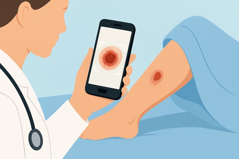
AI models can detect wound boundaries, measure wound area, and estimate tissue types (granulation, slough, necrosis) from digital photos. Automated segmentation reduces manual measurement variability and speeds documentation. Several deep-learning algorithms have been developed and validated on wound-photo datasets and show good agreement with expert measurements in controlled studies. These tools can be embedded in smartphone apps or wound management platforms for clinic and home use.
2. Predictive analytics: forecasting healing and complications
Machine-learning models trained on EHR data, wound-size trends, and comorbidities can predict which wounds are unlikely to heal within a given timeframe (for example, 4–12 weeks) and which patients are at higher risk for adverse outcomes such as infection or amputation. Predictive models can help clinicians triage high-risk patients for early specialist referral or more intensive interventions. Multiple studies show promising predictive accuracy, though models vary by population and data inputs.
3. Decision support for clinicians
AI-based clinical decision support systems (CDSS) can integrate images, lab results, perfusion studies, and patient factors to suggest likely diagnoses (infected vs. colonized wound), flag red-flag features (possible osteomyelitis, ischemia), and recommend evidence-based next steps. Explainable AI (XAI) methods are increasingly used to show clinicians why a model made a suggestion, which helps build trust and supports shared decision-making.
4. Remote monitoring and telewound care
AI paired with telemedicine allows patients or caregivers to upload wound images and sensor data (moisture, temperature, pH). Algorithms can triage images, detect deterioration, and alert clinicians when early intervention is needed. This can reduce unnecessary clinic visits and enable earlier treatment when wounds worsen. IoT-enabled smart sensors and wearable patches are under active development and early clinical testing.
5. Smart dressings and real-time analytics
Next-generation “intelligent” dressings incorporate sensors (temperature, pH, moisture) and can feed continuous data into AI models. These systems aim to detect early signs of infection or increased bioburden before clinical signs appear, enabling preemptive debridement or antibiotic review. Laboratory and early clinical studies demonstrate feasibility; real-world effectiveness depends on robust validation and integration with clinical workflows.
Evidence snapshot (what reviews and trials show)
Image-based AI systems: Multiple studies demonstrate that DL models can segment wounds and estimate area or tissue composition with accuracy comparable to human raters in curated datasets. Performance can drop in real-world photos; external validation matters.
Predictive models: Several ML studies using EHR data have accurately predicted wound healing at 4, 8, and 12 weeks in retrospective cohorts; prospective validation is less common but emerging. Predictive models have been used to flag high-risk patients for intensified management.
Smart dressings and sensors: Research prototypes and small clinical trials report that sensor-equipped dressings can detect changes in temperature or pH that correlate with infection or increased exudate; combining sensor data with AI algorithms improves detection sensitivity. Large multicenter clinical trials remain limited.
Systematic reviews and narrative overviews generally conclude that AI in wound care is promising, particularly for image analysis and risk prediction, but emphasize heterogeneity, potential biases in datasets, and the need for external validation and clinical trials that measure patient-centered outcomes.
Practical benefits clinicians may expect
More consistent documentation (automated wound size and tissue-type measurements).
Earlier triage of patients who need specialist input (vascular, infectious disease, podiatry).
Reduced clinic burden by triaging which images need clinician review versus automated reassurance.
Objective tracking for quality metrics and payer documentation (e.g., percent area reduction over time).
Integration with telehealth to support home-based wound management and reduce travel burden for patients.
Key limitations and implementation challenges
Data quality and generalizability. AI models trained on curated image sets or single-center EHRs may not generalize to new populations, skin tones, camera types, or real-world lighting conditions. External validation across diverse settings is essential.
Bias and equity. Many image datasets under-represent darker skin tones or certain wound types. Bias in training data can reduce performance for some patient groups; developers must prioritize diverse datasets.
Regulatory and safety pathways. AI-based medical devices need regulatory clearance (e.g., FDA, CE) for clinical use. Regulatory expectations include demonstration of safety, performance, and post-market surveillance. Clinicians should confirm clearance and understand intended use.
Explainability and clinician trust. “Black-box” models can be hard to trust. Explainable AI techniques (heatmaps, feature importance) help clinicians understand model outputs and integrate them into care.
Workflow integration. AI tools must fit existing clinical workflows and EHR systems. Poorly integrated solutions add friction and reduce adoption. Workflows should preserve clinician oversight and medico-legal responsibility.
Privacy and security. Wound photos and sensor data are protected health information. Secure transmission, storage, and consent are essential. Developers and health systems must follow HIPAA/GDPR and best practices for cybersecurity.
Ethics, reimbursement, and practice considerations
Ethics: AI should augment, not replace, clinician judgement. Consent for image use, transparency about AI role, and equitable performance across patient groups are ethical priorities.
Reimbursement: Few AI wound tools have direct reimbursement codes yet. In many systems, AI's value will be demonstrated through reduced clinic visits, improved healing rates, or prevented admissions (which could attract payer interest). Health economic studies are emerging.
Training and education: Clinicians need training on interpreting AI outputs and on when to override automated suggestions. Wound-care education should include basic AI literacy so teams can assess vendor claims critically.
Practical adoption checklist for clinicians and clinics
Ask for evidence. Request peer-reviewed validation studies showing performance on external datasets and clinical outcomes.
Check regulatory status. Confirm the product’s clearance/approval for the intended use.
Evaluate integration. Ensure the tool connects with your EHR or workflow and secures PHI.
Pilot locally. Start with a small pilot to evaluate performance, documentation benefit, and clinician acceptance. Collect feedback and measure time saved or changes in triage.
Monitor performance. Track discrepancies between AI outputs and clinician assessments, and report adverse events. Implement an ongoing quality-assurance process.
Address equity. Confirm the vendor tested performance across a diverse range of skin tones and wound types. If gaps exist, proceed cautiously.
Research and future directions
Large multi-center prospective trials that test AI tools as part of clinical workflows and measure patient-centered outcomes (healing rates, time to closure, hospitalization) are needed.
Standardized datasets and benchmarks will help compare algorithms fairly and encourage robust external validation. Open, diverse image repositories are in progress in some academic networks.
Better sensor–AI integration: real-time analytics from smart dressings combined with predictive models may enable earlier detection of infection or ischemia. Early engineering and clinical studies are promising.
Explainability and human–AI teaming: designing interfaces that show the reasoning behind predictions will improve clinician adoption and patient safety.
Takeaway: how to think about AI in wound care today
AI is a promising, practical tool for wound assessment, prediction, and remote monitoring — particularly in image analysis and risk stratification. Early evidence supports improved measurement consistency and potentially earlier triage of high-risk wounds, while smart dressings and sensor-driven AI are exciting but still maturing. Clinicians should seek validated, regulated tools that integrate with their workflows, monitor performance locally, and ensure equitable performance across patient groups. Use AI to augment clinical judgment and to free clinicians for higher-value tasks, while staying cautious about limitations and ensuring patient safety.
See also
When to Use Skin Substitutes or Grafts for Non-Healing Wounds
Stem Cells, Exosomes, and Biologics: Do They Work in Wound Care?
How to Tell If a Wound Is Healing: Signs of Proper Wound Care Progress
Why Diabetic Foot Wounds Heal Slowly: Top Factors That Delay Recovery
How Often Should Wound Dressings Be Changed? Best Practices for Healing
More Information
For more information on the latest effective wound care, contact us to set up a time for a call.
Sources
Berezo M, Budman J, Deutscher D, Hess CT, Smith K, Hayes D. Predicting Chronic Wound Healing Time Using Machine Learning. Advances in Wound Care (New Rochelle). June 2022;11(6):281–96. doi: 10.1089/wound.2021.0073. Epub 2022 Mar 24. PMCID: PMC8982125. https://pmc.ncbi.nlm.nih.gov/articles/PMC8982125/
Rani Raju N, Silina E, Stupin V, Manturova N, Chidambaram SB, Achar RR. Multifunctional and Smart Wound Dressings-A Review on Recent Research Advancements in Skin Regenerative Medicine. Pharmaceutics. 2022 Jul 28;14(8):1574. doi: 10.3390/pharmaceutics14081574. PMID: 36015200; PMCID: PMC9414988. https://pmc.ncbi.nlm.nih.gov/articles/PMC9414988/
Noushin T, Hossain NI and Tabassum S (2022) IoT-Enabled Integrated Smart Wound Sensor for Multiplexed Monitoring of Inflammatory Biomarkers at the Wound Site. Frontiers in Nanotechnoly. 4:851041. doi: 10.3389/fnano.2022.851041. https://www.frontiersin.org/articles/10.3389/fnano.2022.851041/full
Mirani B, Hadisi Z, Pagan E, Dabiri SMH, van Rijt A, Almutairi L, Noshadi I, Armstrong DG, Akbari M. Smart Dual-Sensor Wound Dressing for Monitoring Cutaneous Wounds. Adv Healthc Mater. 2023 Jul;12(18):e2203233. doi: 10.1002/adhm.202203233. Epub 2023 Mar 29. Erratum in: Adv Healthc Mater. 2025 Sep;14(23):e2502264. doi: 10.1002/adhm.202502264. PMID: 36929644; PMCID: PMC11468884. https://onlinelibrary.wiley.com/doi/10.1002/adhm.202203233
https://pmc.ncbi.nlm.nih.gov/articles/PMC11468884/LLe DTP, Pham TD. Unveiling the role of artificial intelligence for wound assessment and wound healing prediction. Explor Med. 2023;4:589–611. https://doi.org/10.37349/emed.2023.00163 https://www.explorationpub.com/Journals/em/Article/1001163
Griffa D, Natale A, Merli Y, Starace M, Curti N, Mussi M, Castellani G, Melandri D, Piraccini BM, Zengarini C. Artificial Intelligence in Wound Care: A Narrative Review of the Currently Available Mobile Apps for Automatic Ulcer Segmentation. BioMedInformatics. 2024; 4(4):2321-2337. https://doi.org/10.3390/biomedinformatics4040126 https://www.mdpi.com/2673-7426/4/4/126
Dallmann AC, Sheridan M, Mattke S, Ennis W. Prediction of Healing Trajectory of Chronic Wounds Using a Machine Learning Approach. Adv Wound Care (New Rochelle). 2024 Nov 6. doi: 10.1089/wound.2024.0095. Epub ahead of print. PMID: 39508072. https://www.liebertpub.com/doi/10.1089/wound.2024.0095
https://pubmed.ncbi.nlm.nih.gov/39508072/Rathore, P.S., Kumar, A., Nandal, A. et al. A feature explainability-based deep learning technique for diabetic foot ulcer identification. Sci Rep 15, 6758 (2025). https://doi.org/10.1038/s41598-025-90780-z https://www.nature.com/articles/s41598-025-90780-z
Teagan Weatherall, Pinar Avsar, Linda Nugent, Zena Moore, John H. McDermott, Seamus Sreenan, Hannah Wilson, Natalie L. McEvoy, Rosemarie Derwin, Paul Chadwick, Declan Patton, The impact of machine learning on the prediction of diabetic foot ulcers – A systematic review, Journal of Tissue Viability, Volume 33, Issue 4, 2024, Pages 853-863, ISSN 0965-206X, https://doi.org/10.1016/j.jtv.2024.07.004 https://www.sciencedirect.com/science/article/pii/S0965206X24001098
Su R, Wang L, Han F, Bian S, Meng F, Qi W, Zhai X, Li H, Wu J, Pan X, Pan H, Guo P, Lu WW, Liu Z, Zhao X. A highly stretchable smart dressing for wound infection monitoring and treatment. Mater Today Bio. 2024 May 31;26:101107. doi: 10.1016/j.mtbio.2024.101107. PMID: 38952538; PMCID: PMC11216007. https://pmc.ncbi.nlm.nih.gov/articles/PMC11216007/
* This blog is for informational purposes only and is not a substitute for professional medical advice, diagnosis, or treatment.