Early Warning Signs of Osteomyelitis in Foot Wounds: What to Look For
Learn the early warning signs of osteomyelitis in foot wounds, including key symptoms, tests, and best practices for early detection and wound care.
admin
10/18/20258 min read
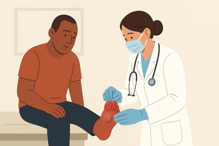
Osteomyelitis, infection of the bone, is a serious complication that can arise from a foot wound. It is especially important to detect early in people with diabetes, peripheral vascular disease, or compromised immune systems because delayed diagnosis may lead to more invasive treatments, prolonged antibiotics, or limb-threatening outcomes. This guide explains the early warning signs of osteomyelitis in foot wounds, practical bedside checks, useful tests, and when to escalate care.
Why early detection matters
Bone infection can be harder to treat than surface infection because bone is relatively avascular and bacteria can hide in biofilm. Early recognition helps clinicians obtain cultures, plan targeted therapy, and decide whether surgical debridement or revascularization is needed. Detecting osteomyelitis early may reduce the duration of antibiotic therapy and lower the risk of complications. That said, diagnosing osteomyelitis can be challenging and usually requires a mix of clinical assessment, imaging, laboratory tests, and sometimes bone sampling.
The clinical picture: symptoms and signs to watch for
No single symptom proves osteomyelitis, but certain patterns raise the index of suspicion. Below are early warning signs grouped into local and systemic features.
Local signs that may suggest bone involvement
Wound failing to heal as expected: an ulcer that has been present for many weeks (commonly >6 weeks) or one that stalls despite appropriate wound care (debridement, offloading, dressings). Chronic nonhealing can be a red flag.
Visible or palpable bone: any wound that shows exposed bone or where blunt probing reaches bone increases the likelihood of underlying osteomyelitis. A positive probe-to-bone (PTB) test is a notable clinical clue.
New or expanding deep infection: increasing depth, a draining sinus tract that tracks toward bone, or a wound that previously looked superficial but becomes deeper.
Red, swollen or “sausage” toe: in toes, a red, hot, swollen digit (sometimes called a “sausage toe”) can be an early sign that infection has reached deeper tissues and possibly bone.
Persistent or increasing purulent drainage or malodour despite standard care; especially if drainage becomes frankly purulent or changes in character.
Systemic or subtler signs
Low-grade systemic inflammation: mild fever is possible but not always present. Many patients (especially older adults or those with diabetes) may lack robust systemic signs.
Laboratory markers: elevated inflammatory markers such as erythrocyte sedimentation rate (ESR) or C-reactive protein (CRP) can support suspicion. These tests are not definitive alone but can help when combined with other findings. ESR often shows higher sensitivity for osteomyelitis than other markers.
Useful bedside tests and what their results mean
Probe-to-bone (PTB) test
The PTB test is a simple maneuver: after cleaning the wound, a sterile blunt probe is used to see if bone can be felt at the base. Systematic reviews suggest PTB has reasonable diagnostic value. It tends to perform better in settings where the prevalence of osteomyelitis is higher (for example, in referral clinics for diabetic foot ulcers). A positive PTB increases the probability of bone infection; a negative PTB does not completely rule it out. Use PTB as part of the assessment rather than a standalone test.
Plain X-ray (radiograph)
X-rays are widely available and may show cortical erosion, periosteal reaction, or bone destruction, but these changes can take time to appear (weeks). A normal X-ray does not exclude early osteomyelitis. However, an abnormal X-ray in the right clinical setting is a helpful clue. Combining PTB and X-ray can improve diagnostic confidence in some cases.
Inflammatory blood tests (ESR, CRP, WBC)
ESR and CRP are commonly used. ESR may be particularly helpful as a screening tool for diabetic foot osteomyelitis; CRP is useful for monitoring response to therapy. Neither test is perfectly sensitive or specific on its own and should be interpreted in context.
Imaging: choosing the right test when you suspect osteomyelitis
When clinical suspicion is moderate to high or initial tests are inconclusive, imaging becomes important.
Magnetic resonance imaging (MRI)
MRI is generally considered the most sensitive imaging for early osteomyelitis and for assessing the extent of bone and soft-tissue involvement. MRI can detect bone marrow edema and adjacent abscesses earlier than X-ray. However, MRI may be less available in some settings and can be limited by implants or severe renal impairment (contrast considerations). If feasible, MRI is often the best next step when osteomyelitis is suspected.
Nuclear medicine scans
Techniques such as labeled white blood cell (WBC) scans, Technetium bone scans, or combined WBC + sulfur colloid imaging may be used when MRI is contraindicated or unavailable. These tests have variable performance characteristics and are often complementary to MRI.
Ultrasound and CT
Ultrasound may identify soft-tissue abscesses or periosteal changes in some scenarios but is less sensitive for marrow involvement. CT can show cortical bone destruction and can be useful for surgical planning or when MRI is not an option.
Microbiology: how to get the right specimen
If osteomyelitis is suspected, obtaining a microbiological diagnosis helps target antibiotics. Best practice is to obtain deep tissue or bone samples (not superficial swabs) after appropriate debridement or during surgery. If surgery is not feasible, percutaneous image-guided bone biopsy may be considered. Superficial swabs often reflect surface colonizers and are of limited value for guiding systemic therapy for bone infection.
Typical clinical scenarios that should raise concern
A diabetic patient with a plantar forefoot ulcer that has been present for >6 weeks, especially if callus or recurrent breakdown is present.
A wound with exposed bone or a sinus tract that probes to bone.
A previously stable chronic wound that acutely becomes more painful, red, or draining purulence.
A “sausage toe” or red, swollen digit in a patient with neuropathy or diabetes.
In any of these scenarios, consider prompt imaging (often MRI), blood tests (ESR/CRP), and urgent discussion with the multidisciplinary team (wound care, infectious diseases, surgery/podiatry, and vascular if ischemia is suspected).
Early management steps while you investigate
Optimize local wound care: debridement of devitalized tissue, appropriate dressings, and offloading for plantar wounds. Mechanical disruption of biofilm is often an important first step.
Obtain blood tests: ESR and CRP can help gauge the inflammatory response and monitor progress.
Perform PTB and X-ray at the bedside as immediate checks; if positive or if clinical suspicion remains, arrange MRI or specialist imaging.
Avoid premature systemic antibiotics if the diagnosis is uncertain and bone sampling is planned; preoperative antibiotics may obscure culture results. If systemic infection or sepsis is suspected, start empiric antibiotics and obtain cultures promptly.
When to involve specialists and when to refer urgently
Urgent surgical review is needed for suspected deep abscess, rapidly spreading infection, or systemic toxicity.
Podiatry/orthopedics for suspected osteomyelitis that may require bone biopsy or surgical debridement/resection.
Infectious disease for guidance on antibiotic choices and durations, particularly for confirmed osteomyelitis.
Vascular surgery if ischemia or poor perfusion is suspected, since revascularization can be important for healing and antibiotic delivery.
Early multidisciplinary input can speed diagnosis and improve outcomes.
Putting it together: a simple checklist for clinicians
When evaluating a foot wound for possible osteomyelitis, ask:
Is there exposed bone or does the PTB test reach bone?
Has the ulcer been present >6 weeks or stalled despite care?
Is there increasing depth, purulence, sinus tract, or a red/swollen toe?
Are ESR/CRP elevated in a compatible clinical picture?
Do X-ray or MRI findings suggest bone involvement?
Can we obtain deep tissue or bone cultures before starting antibiotics (if safe)?
Have we engaged the multidisciplinary team (surgery, ID, vascular) as needed?
If several answers are “yes,” act promptly: arrange MRI if possible, obtain appropriate cultures, and involve specialists for potential surgical and antibiotic planning.
Limitations
No single symptom, sign, or test definitively diagnoses osteomyelitis in isolation. Many chronic wounds are colonized with bacteria without bone invasion. Diagnostic accuracy depends on the clinical setting, pretest probability, and the quality of imaging and sampling. The best approach combines careful bedside assessment, appropriate imaging, laboratory data, and, when possible, microbiological confirmation from deep samples.
Final thoughts
Early recognition of osteomyelitis in foot wounds requires vigilance. Watch for wounds that fail to progress, exposed bone or a positive probe-to-bone test, increasing local signs (redness, swelling, purulent drainage), and supportive lab or imaging findings. When suspicion is moderate to high, prioritize MRI and bone sampling where feasible, and engage a multidisciplinary team. Acting early — while balancing the need for accurate culture-guided therapy — can help avoid prolonged infection, reduce antibiotic misuse, and support better healing.
See also
When to Use Systemic Antibiotics in Wound Care: Best Practices
Wound Care Guide: How to Tell Colonization from True Infection
How to Tell If a Wound Is Healing: Signs of Proper Wound Care Progress
Why Diabetic Foot Wounds Heal Slowly: Top Factors That Delay Recovery
How Often Should Wound Dressings Be Changed? Best Practices for Healing
More Information
For more information on the latest effective wound care, contact us to set up a time for a call.
Sources
Senneville E, et al. 2023. Guidelines on the diagnosis and treatment of foot infection in persons with diabetes. International Working Group on the Diabetic Foot (IWGDF/IDSA).
https://iwgdfguidelines.org/wp-content/uploads/2023/07/IWGDF-2023-04-Infection-Guideline.pdfKenrick Lam, Suzanne A. V. van Asten, Tea Nguyen, Javier La Fontaine, Lawrence A. Lavery, Diagnostic Accuracy of Probe to Bone to Detect Osteomyelitis in the Diabetic Foot: A Systematic Review, Clinical Infectious Diseases, Volume 63, Issue 7, 1 October 2016, Pages 944–948, https://doi.org/10.1093/cid/ciw445
https://academic.oup.com/cid/article/63/7/944/2197008Woo I, Cho SJ, Park CH. State-of-the-art update for diagnosing diabetic foot osteomyelitis: a narrative review. Journal of Yeungnam Medical Science. 2023;40(4):321-327.
https://e-jyms.org/journal/view.php?doi=10.12701/jyms.2023.00976Mutluoglu M, Lipsky BA. Diabetic foot osteomyelitis. Canadian Medical Association Journal. 2016 Dec 6;188(17-18):E535. doi: 10.1503/cmaj.160228. Epub 2016 Oct 17. PMID: 27754891; PMCID: PMC5135537.
https://www.cmaj.ca/content/188/17-18/E535Rosario Morales Lozano, Maria L. González Fernández, David Martinez Hernández, Juan V. Beneit Montesinos, Sagrario Guisado Jiménez, Maximo A. Gonzalez Jurado; Validating the Probe-to-Bone Test and Other Tests for Diagnosing Chronic Osteomyelitis in the Diabetic Foot. Diabetes Care 1 October 2010; 33 (10): 2140–2145. https://doi.org/10.2337/dc09-2309 https://diabetesjournals.org/care/article/33/10/2140/28212/Validating-the-Probe-to-Bone-Test-and-Other-Tests
Calvo-Wright MdM, Álvaro-Afonso FJ, López-Moral M, García-Álvarez Y, García-Morales E, Lázaro-Martínez JL. Is the Combination of Plain X-ray and Probe-to-Bone Test Useful for Diagnosing Diabetic Foot Osteomyelitis? A Systematic Review and Meta-Analysis. Journal of Clinical Medicine. 2023; 12(16):5369. https://doi.org/10.3390/jcm12165369. https://pmc.ncbi.nlm.nih.gov/articles/PMC10455253/
Momodu II, Savaliya V. Osteomyelitis. [Updated 2023 May 31]. In: StatPearls [Internet]. Treasure Island (FL): StatPearls Publishing; 2025 Jan-. Available from: https://www.ncbi.nlm.nih.gov/books/NBK532250/
Anurag Markanday, Diagnosing Diabetic Foot Osteomyelitis: Narrative Review and a Suggested 2-Step Score-Based Diagnostic Pathway for Clinicians, Open Forum Infectious Diseases, Volume 1, Issue 2, Summer 2014, ofu060, https://doi.org/10.1093/ofid/ofu060
https://academic.oup.com/ofid/article/1/2/ofu060/1465780Mayo Clinic — Osteomyelitis patient-friendly overview. https://www.mayoclinic.org/diseases-conditions/osteomyelitis/symptoms-causes/syc-20375913
McCreery R, et al. Diabetes-Related Foot Infections: Institutional Treatment Guidance. University of Nebraska Medical Center.
https://www.unmc.edu/intmed/_documents/id/asp/clinicalpath-dfi_institutional_guideline_final.pdfACR Appropriateness Criteria - Suspected Osteomyelitis of the Foot. 2025. American College of Radiology.
https://acsearch.acr.org/docs/69340/NarrativeSharma H, Sharma S, Krishnan A, Yuan D, Vangaveti VN, Malabu UH, et al. (2022) The efficacy of inflammatory markers in diagnosing infected diabetic foot ulcers and diabetic foot osteomyelitis: Systematic review and meta-analysis. PLoS ONE 17(4): e0267412. https://doi.org/10.1371/journal.pone.0267412 https://journals.plos.org/plosone/article?id=10.1371/journal.pone.0267412
2019. Osteomyelitis. National Organization for Rare Disorders.
https://rarediseases.org/rare-diseases/osteomyelitis/Schaper N, et al. 2023. Practical guidelines on the prevention and management of diabetes-related foot disease. International Working Group on the Diabetic Foot (IWGDF)
https://iwgdfguidelines.org/wp-content/uploads/2023/07/IWGDF-2023-01-Practical-Guidelines.pdf .Ansert EA, et al. Update of biomarkers to diagnose diabetic foot osteomyelitis: A meta-analysis and systematic review. Wound Repair and Regeneration. 2024.
https://pubmed.ncbi.nlm.nih.gov/38566503/Patel D. Osteomyelitis Clinical Presentation. Medscape. 2022.
https://emedicine.medscape.com/article/1348767-clinicalLiu L, Tao Y, Zhang D, Hou J, Zhou G and Tian M (2025) Diagnostic value and integrated threshold of ESR for diabetic foot osteomyelitis: a systemic review and meta-analysis. Frontiers. Endocrinology. 16:1660465. doi: 10.3389/fendo.2025.1660465
https://www.frontiersin.org/journals/endocrinology/articles/10.3389/fendo.2025.1660465/full
* This blog is for informational purposes only and is not a substitute for professional medical advice, diagnosis, or treatment.